一 实验准备
如图1.1所示,搭设3D-DIC系统测量(50mm镜头,2000万像素相机)。

1.1 测量系统示意图
如图1.2所示,使用小型高压喷枪在物体表面喷涂荧光微液滴,喷涂3-5秒(量极小)。需要注意的是,需要提前将组织表面的水分吸干,否则荧光液滴会在湿润的组织表面晕染,无法形成清晰的散斑颗粒。散斑颗粒直径约在50-200微米不等。在紫外光源激发下,试样如图1.3所示。用镊子夹住试样的一角,做一次简单的拉伸,如图1.4。
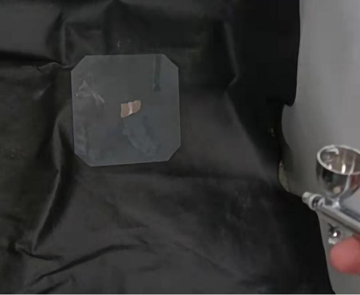
1.2 高压喷枪喷涂荧光散斑
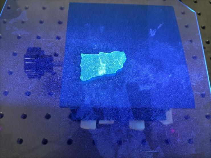
1.3 在紫外光源下激发的荧光散斑
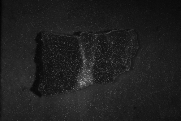
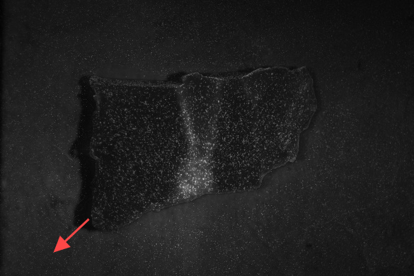
1.4 将组织朝左下角拉伸
二 实验结果
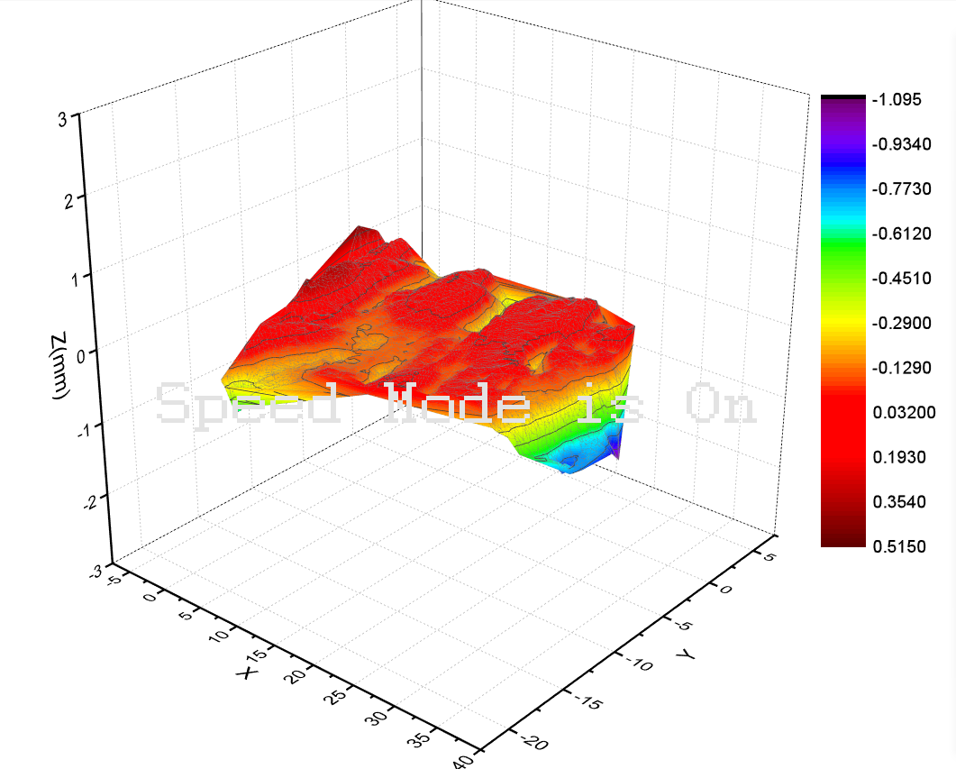
2.1 被测组织表面三维形貌图
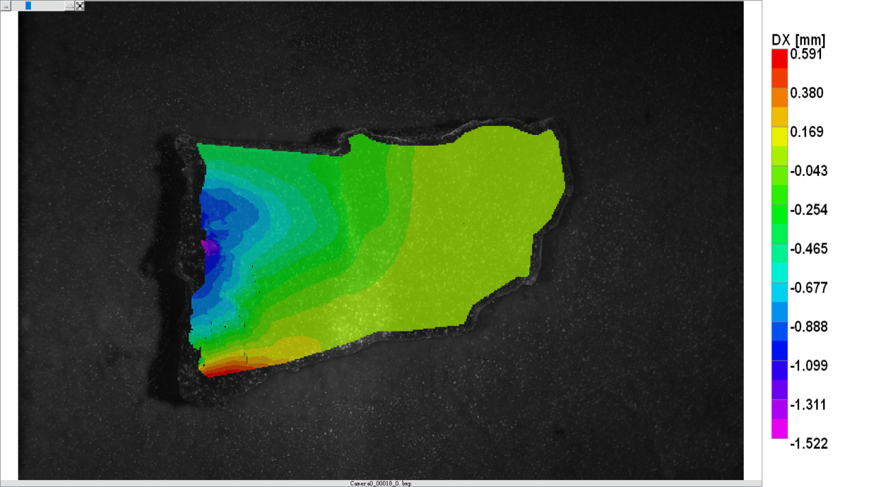
2.2 水平方向位移云图
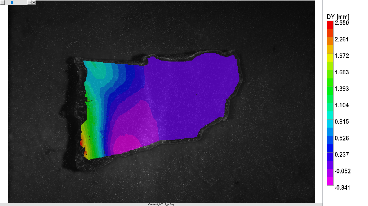
2.3 竖直方向位移云图
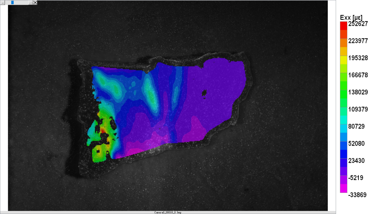
2.4 水平方向应变分布云图
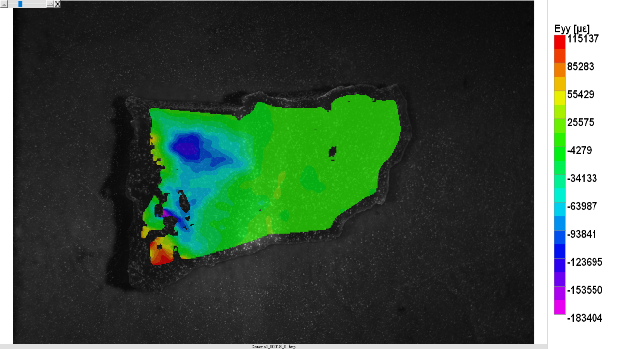
2.5 竖直方向应变分布云图